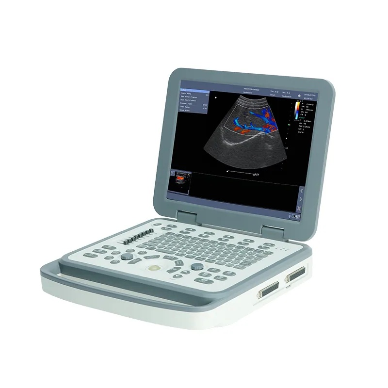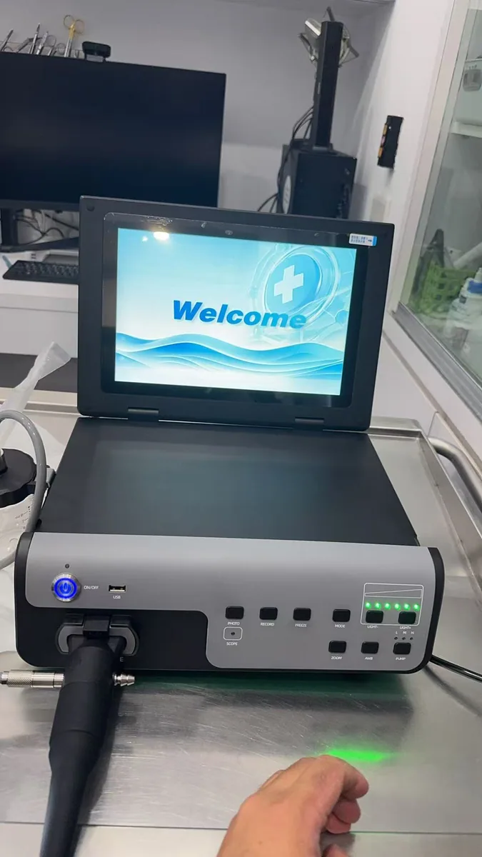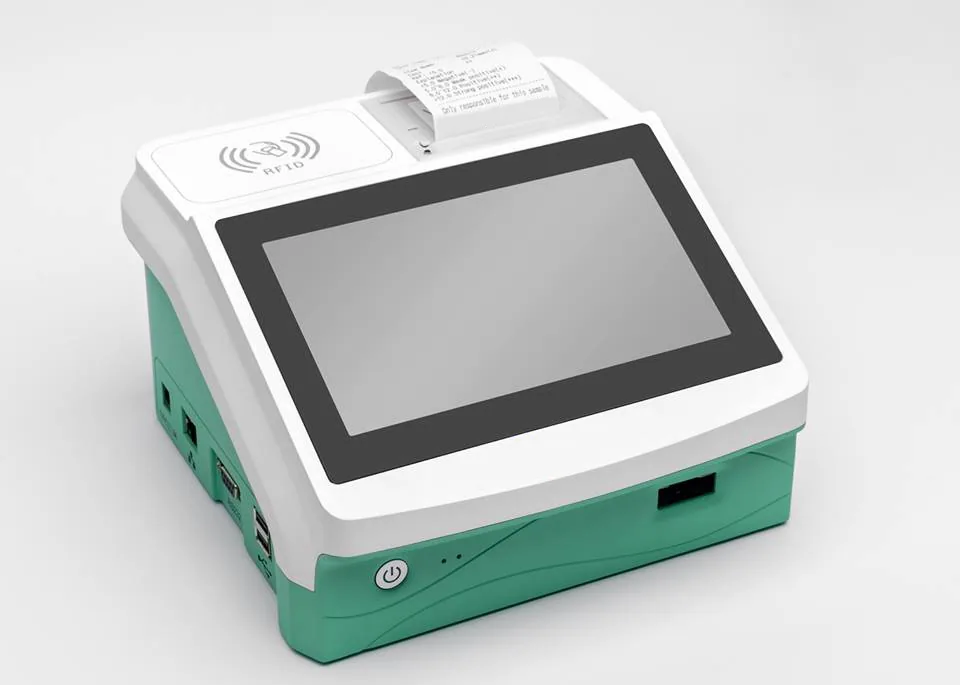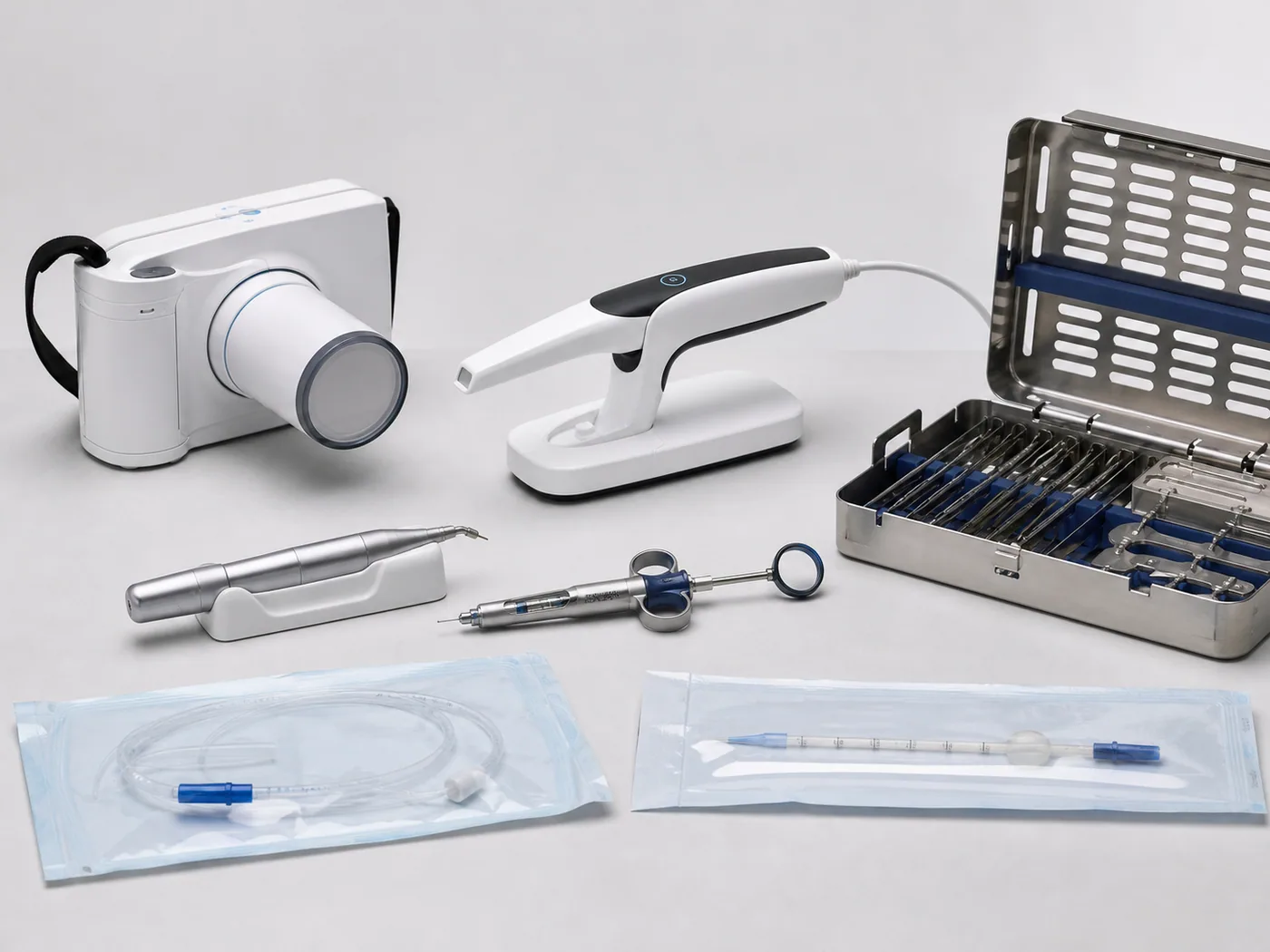
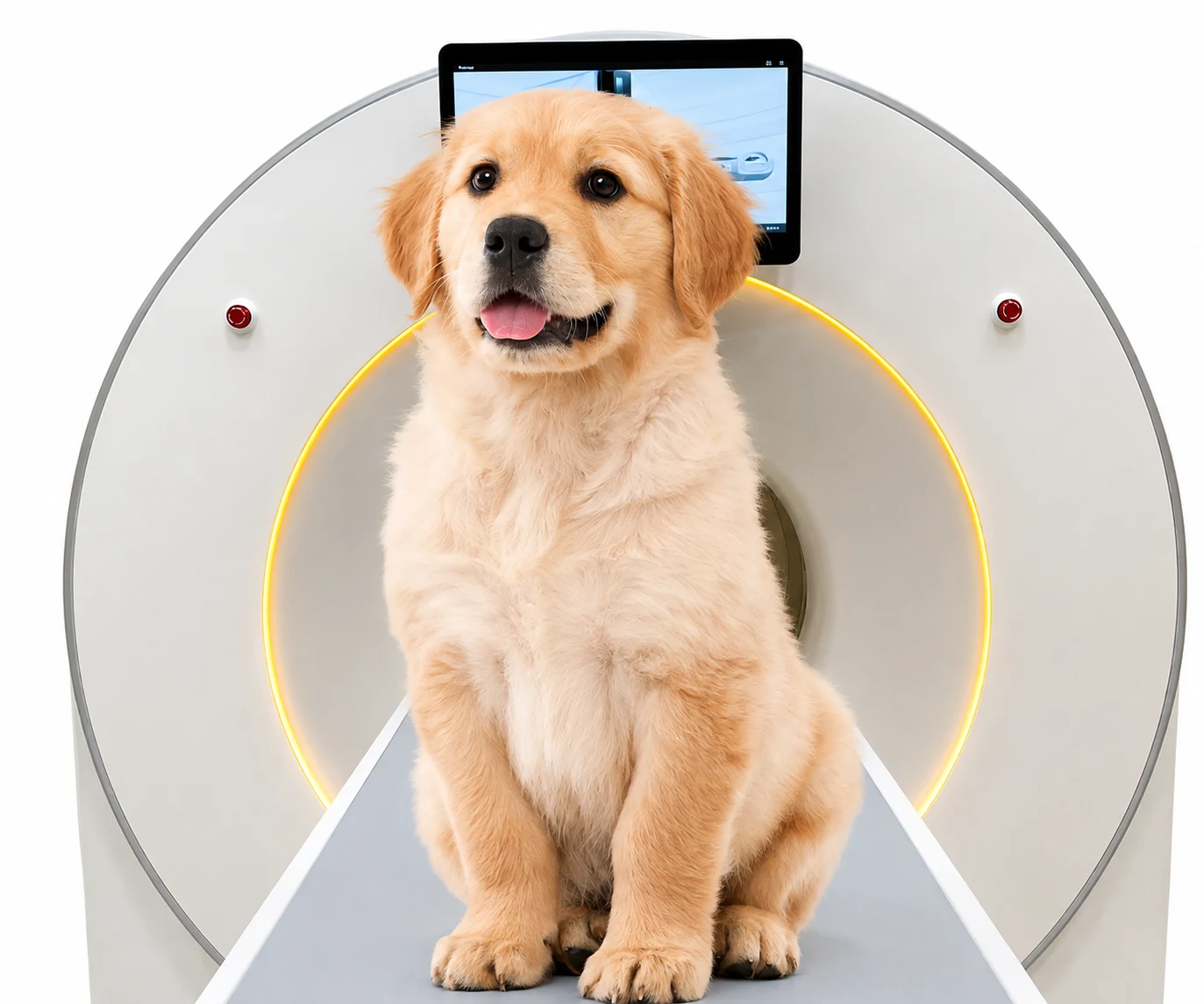
 Supplied product image
01 · MS-P3V
Orthopedic Veterinary CT Scanner
Orthopedic veterinary CT delivers high-resolution 3D bone imaging for precise fracture assessment, joint evaluation and surgical planning.
01 · Banner
Veterinary CT Scanner for Animal Hospitals | Small Animal & Equine CT Imaging
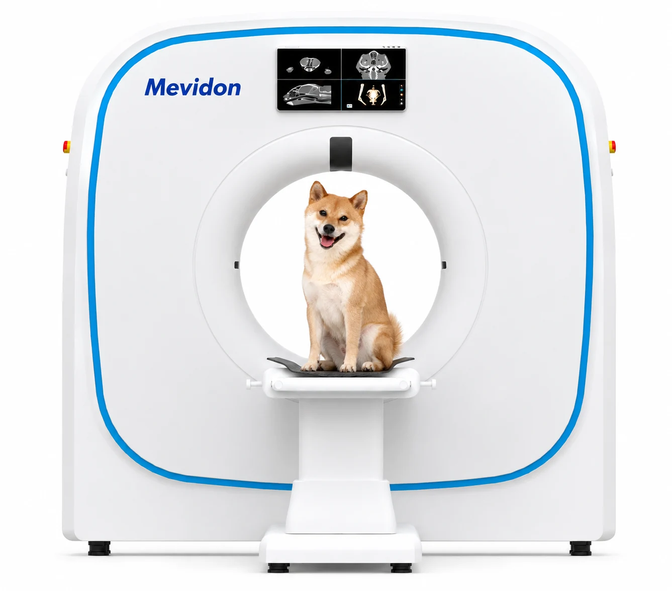
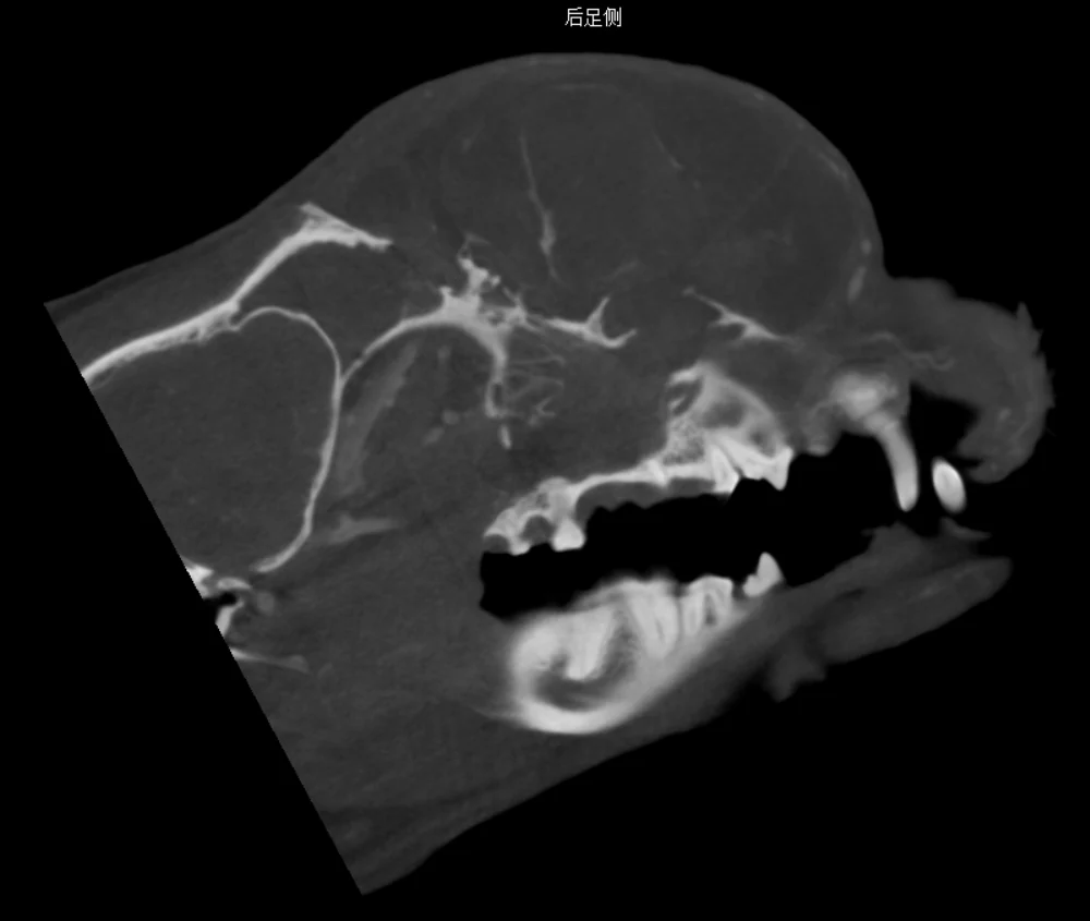
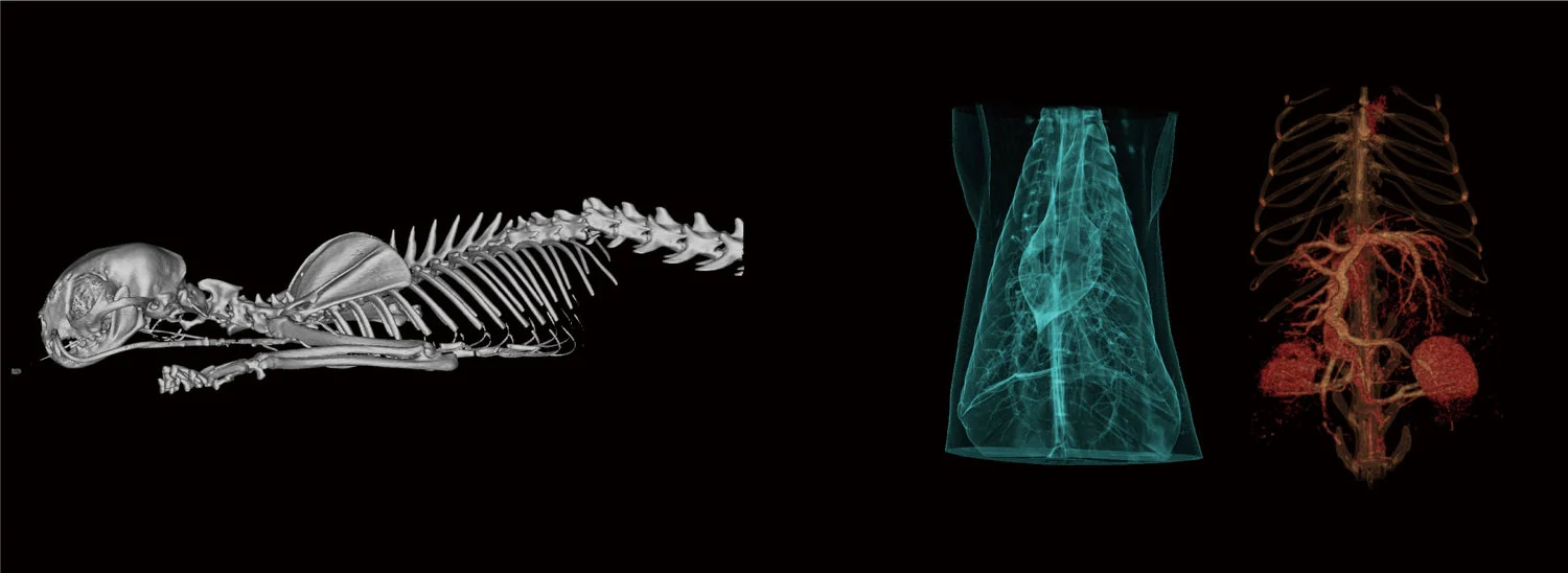 Veterinary CT imaging output
02 · Product Details
03 · Product Descriptions
Veterinary CT scanners help veterinarians see internal structures in greater detail by creating cross-sectional images of the animal body. They are widely used for complex cases where standard radiography or ultrasound may not provide enough anatomical information.
Whether you need a CT scanner for small-animal diagnosis, equine head imaging, dental CT, orthopedic evaluation or tumor staging, our veterinary CT systems support clear image acquisition, accurate anatomical assessment and an efficient clinical workflow.
Mevidon supplies affordable veterinary CT scanners for dental, orthopedic, neurology, oncology, surgery, trauma and maxillofacial applications. CT can provide strong evidence for cancer and tumor staging, metastasis evaluation, biopsy planning and radiation therapy planning. Our systems combine high-resolution image quality, stable performance and responsive after-sales service.
Welcome to send an inquiry for more details.
Send an Inquiry →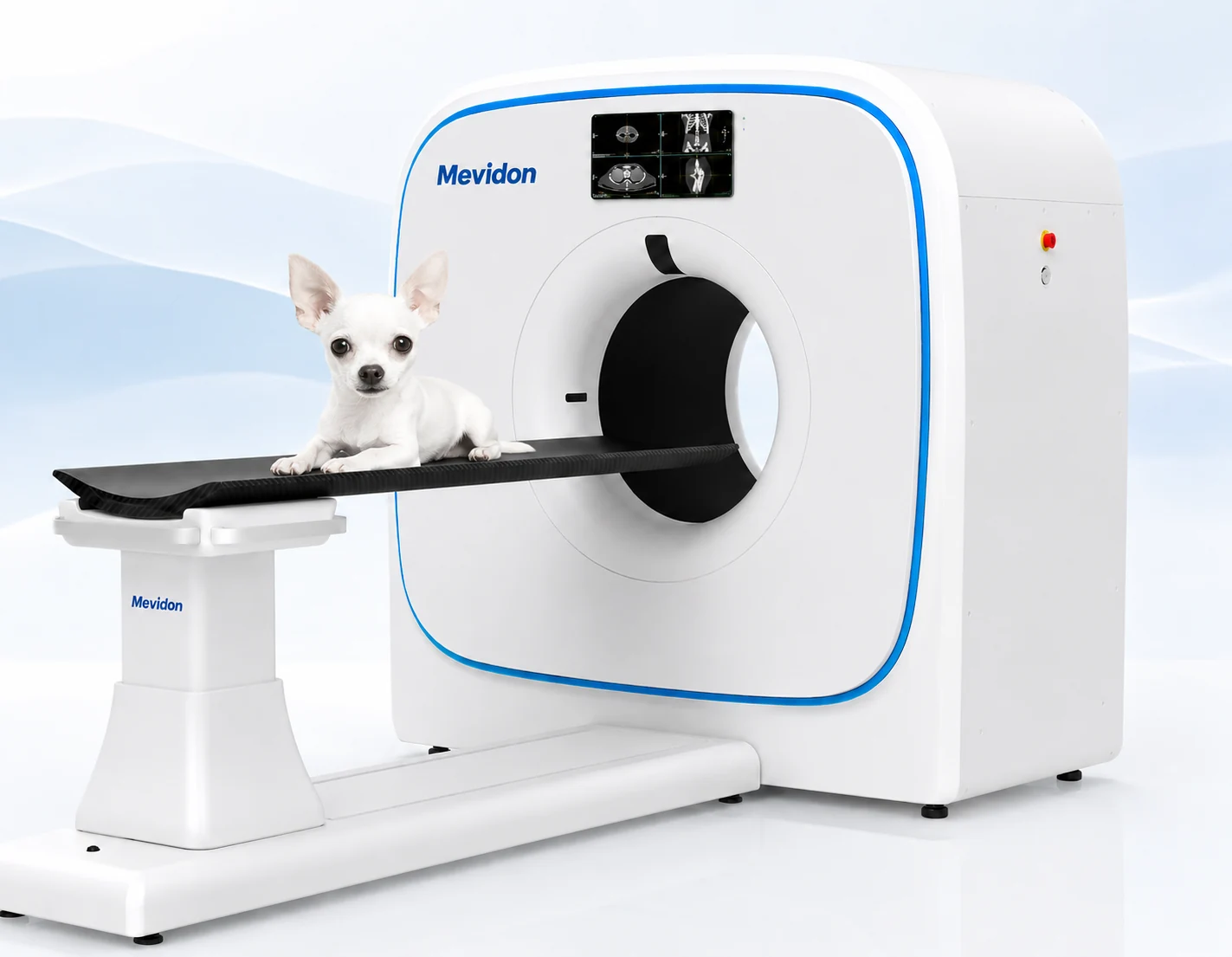 MS-P5V · Supplied product image
04 · Veterinary CT Scanner by Features
Six supplied CT directions for orthopedic, oncology, large-animal, mobile, portable and low-dose veterinary imaging.
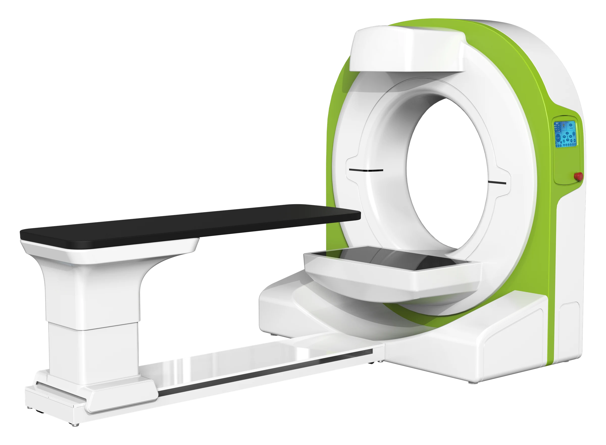
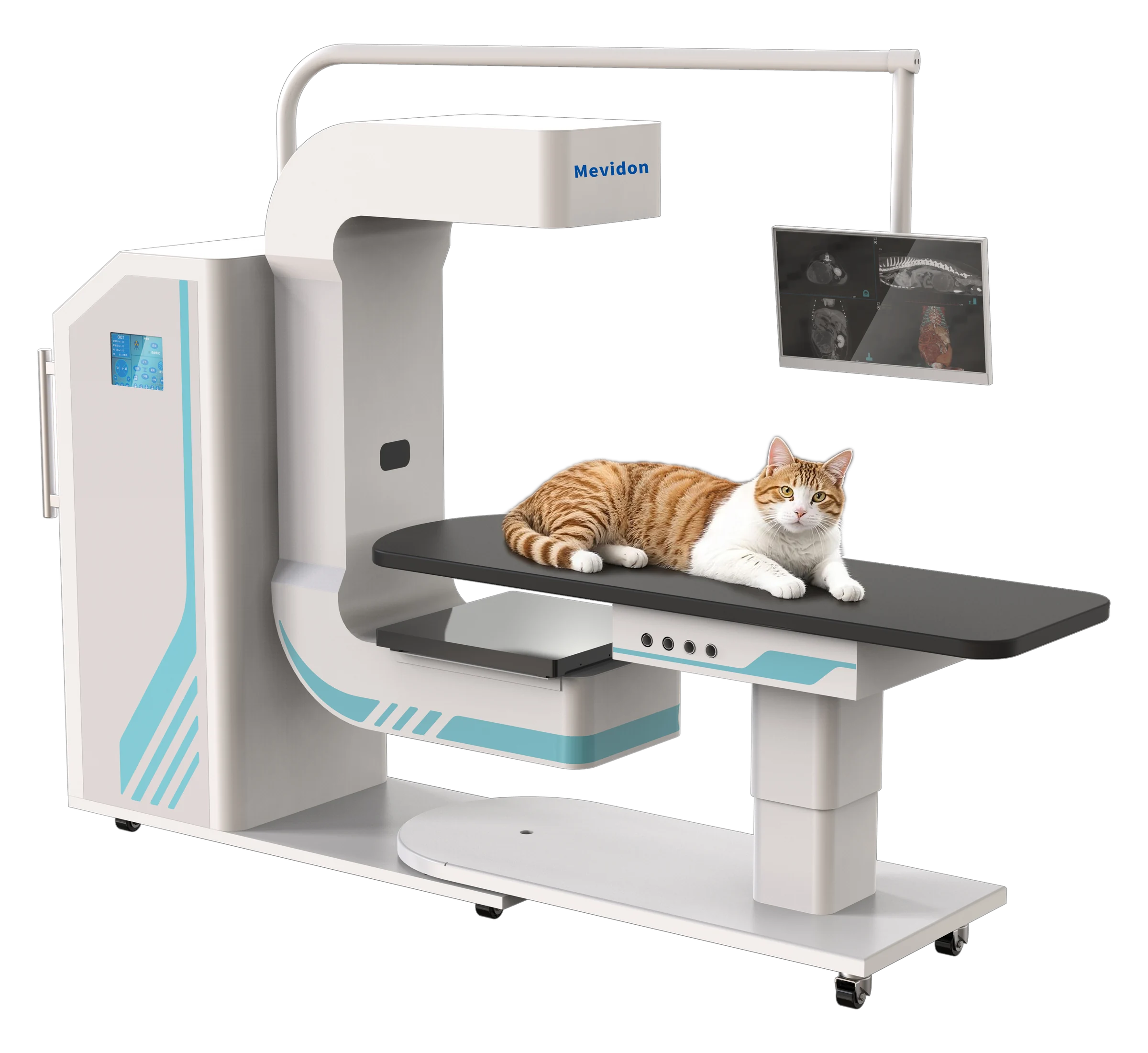
Supplied product imageOrthopedic veterinary CT delivers high-resolution 3D bone imaging for precise fracture assessment, joint evaluation and surgical planning.
 Supplied product image
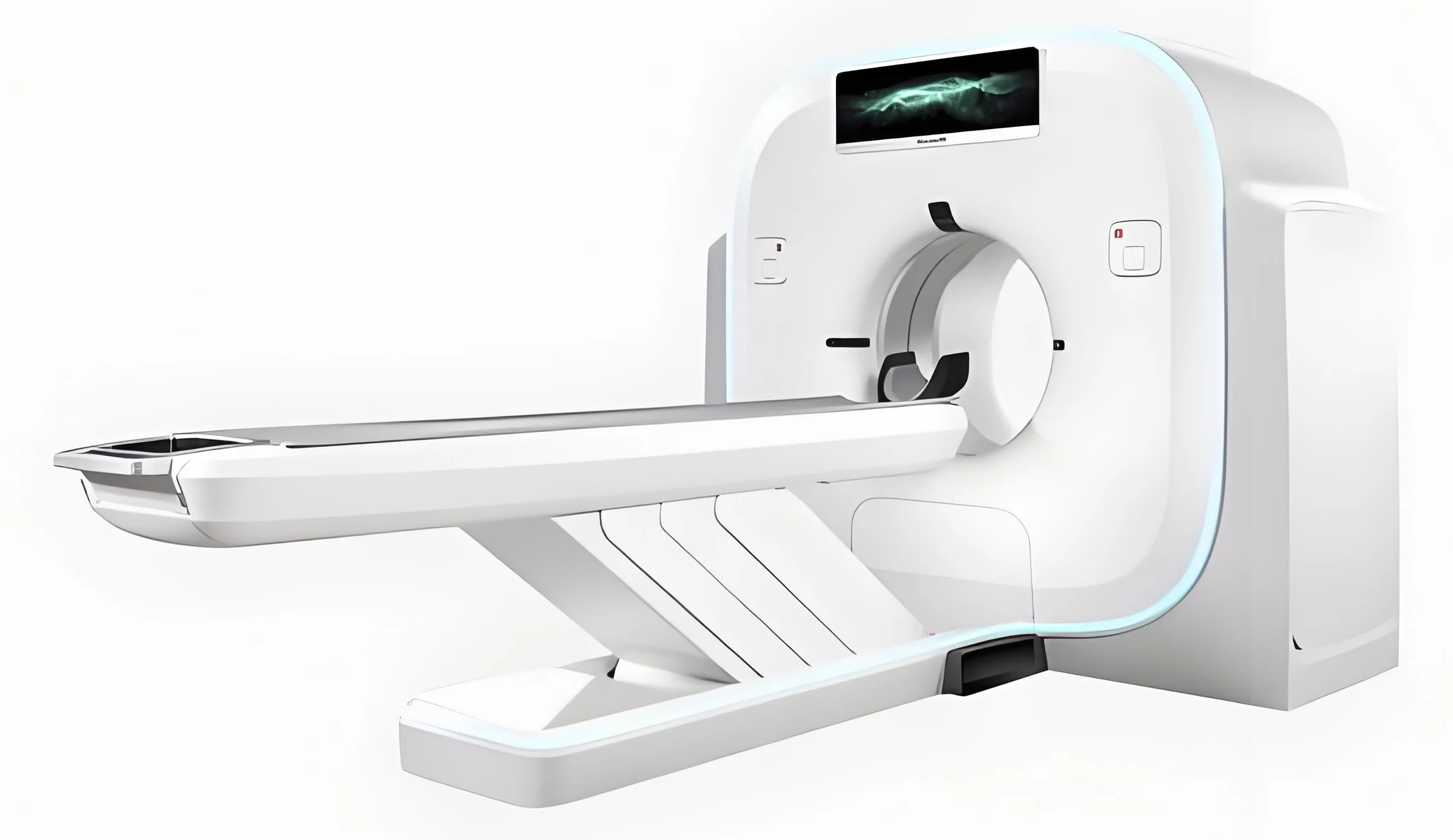
Advanced tumor imaging supports precise cancer staging, metastasis detection, biopsy planning and radiation therapy planning in veterinary oncology.
 Supplied product image
Large-bore CT accommodates horses and large animals, enabling detailed imaging of limbs, spine, pelvis, head and complex anatomy.
 Supplied product image
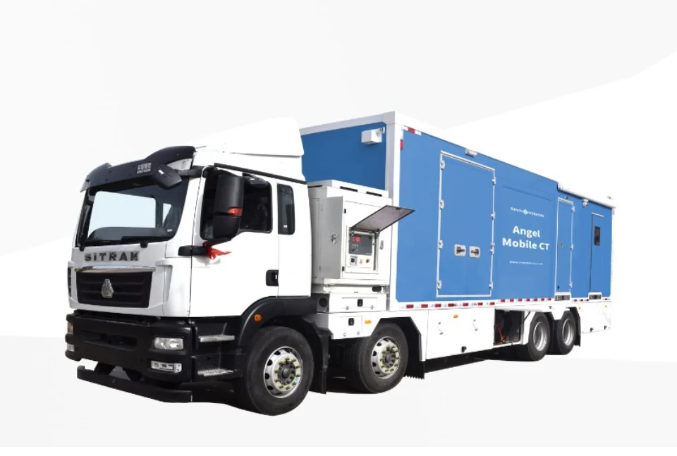
Mobile CT brings advanced cross-sectional imaging closer to veterinary patients, improving access for emergency, surgical and point-of-care diagnostics.
 Supplied product image
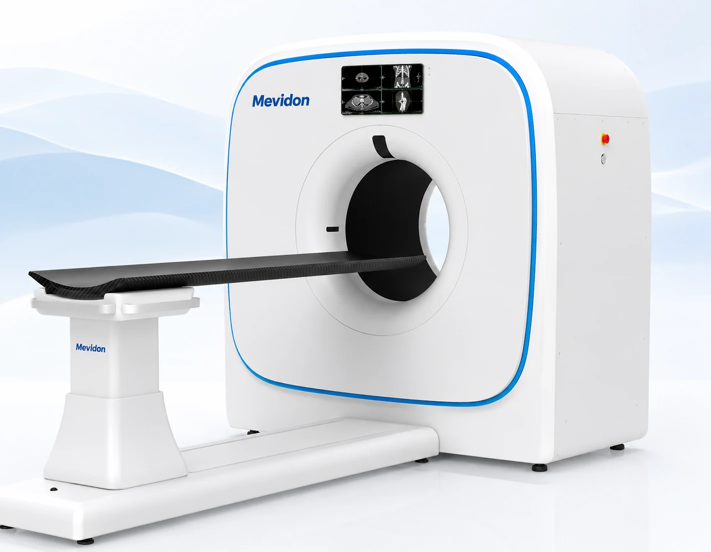
Compact portable CT combines flexible installation with rapid 3D imaging, making advanced diagnostics accessible in space-limited veterinary clinics.
 Supplied product image
Dose-optimized CT reduces radiation exposure while preserving diagnostic image quality for safer repeat examinations and routine veterinary imaging.
05 · Diagnosis of Cancer
Compared with veterinary DR and X-ray, a veterinary CT scanner is suitable for more complex cases. Animal CT is often used for cancer diagnosis because the images can help confirm the presence of tumors, measure tumor size, locate lesions accurately and show their relationship with surrounding tissues. Mevidon animal CT scanners cover dental, orthopedic, oncology, neurology and other scenarios, supporting high-end imaging and multidisciplinary diagnosis.
06 · Company Advantages
Our one-stop animal medical solutions meet different customer needs.

We have a strict quality-control system from procurement and production through outbound inspection.
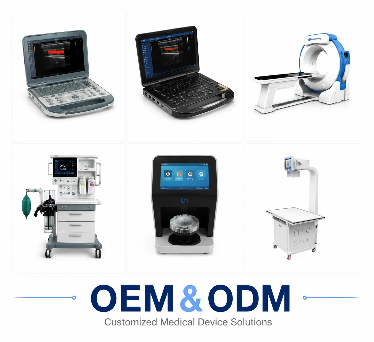
We provide customized OEM and ODM services to design and produce products according to your needs.

More than 1,000 users from pet hospitals and farms choose our veterinary ultrasound machines.
07 · Why Choose Us?
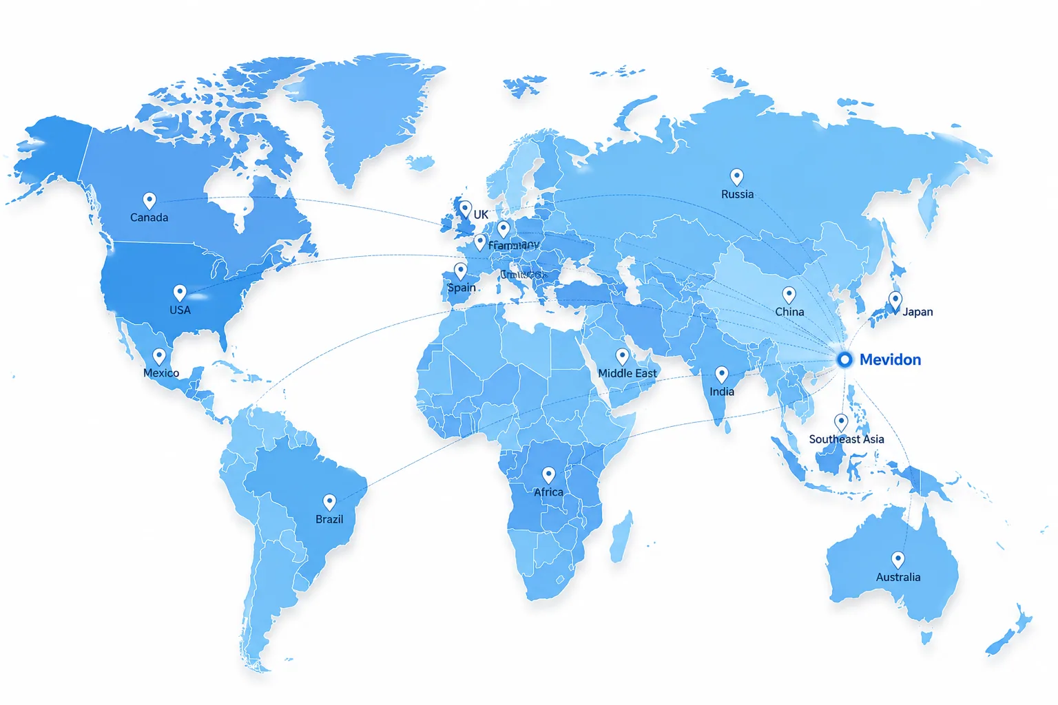
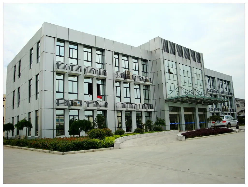
Mevidon has supplied medical and veterinary equipment solutions worldwide. Our customers come from Europe, South America, Latin America, the Middle East, and other regions.

We have rich export experience and are familiar with customs declaration and clearance processes. Professional packaging and complete supply-chain support help us deliver quickly.
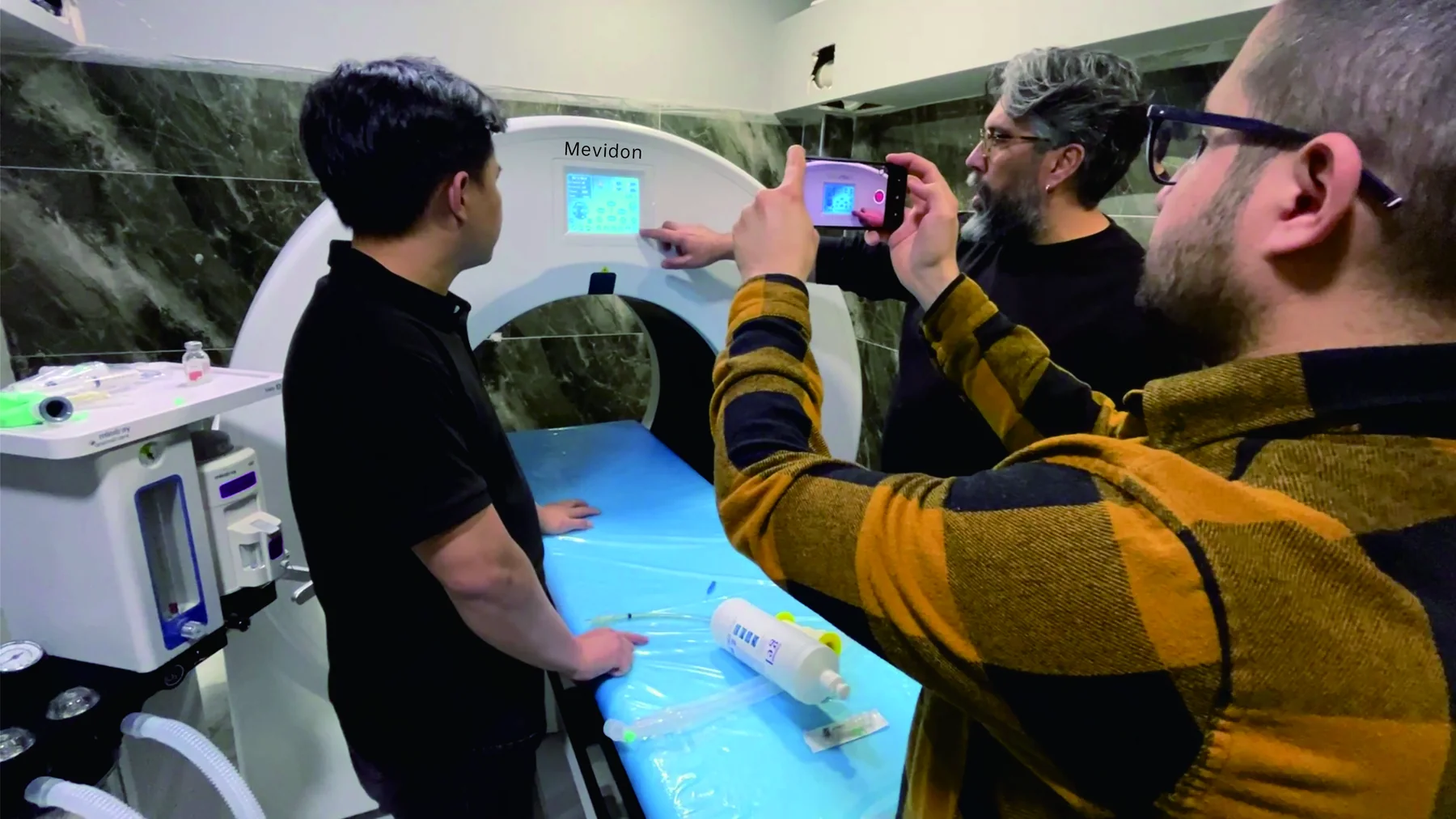
Mevidon provides professional technical training, remote support, operational guidance, online upgrades, and a response within 24 hours.
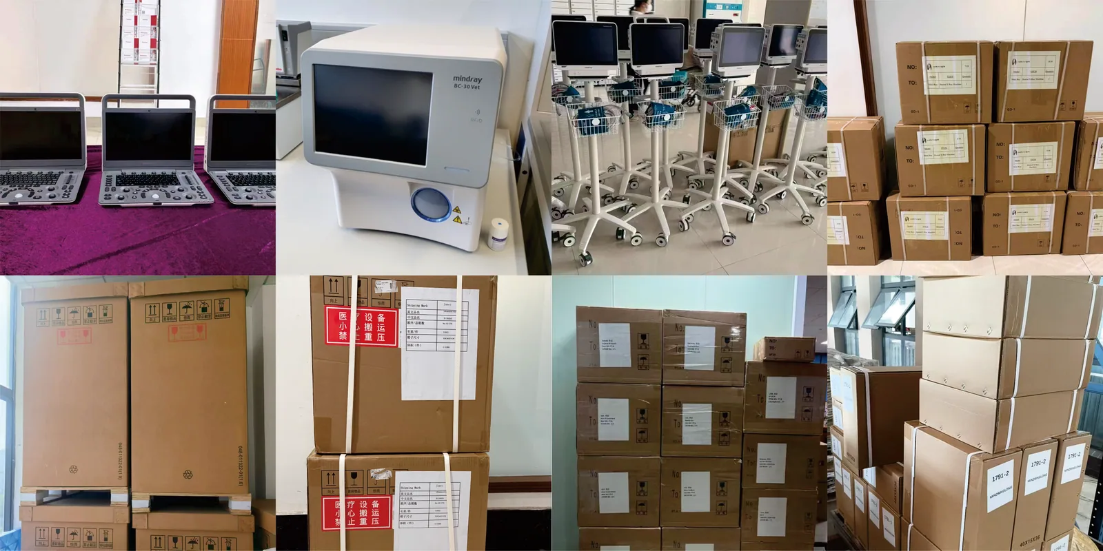
Our animal medical equipment is exported worldwide, and more and more customers choose to cooperate with us.
08 · Clinical Applications
Authentic clinical and workflow materials supplied for this veterinary CT collection.
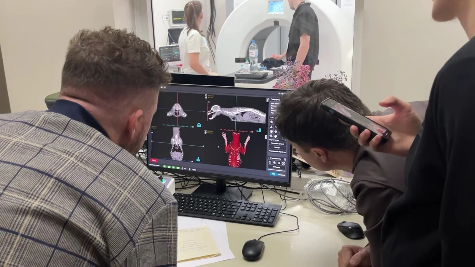
Veterinary CT scanners are widely used in animal hospitals for cats, dogs and other patients.
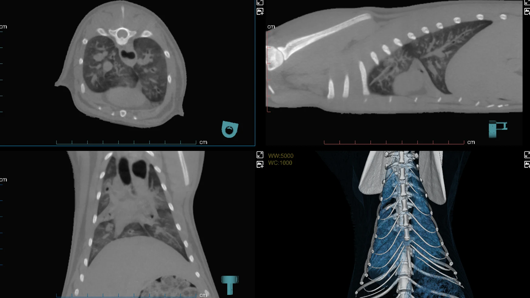
Diagnostic images of lung inflammation acquired with an animal CT scanner.
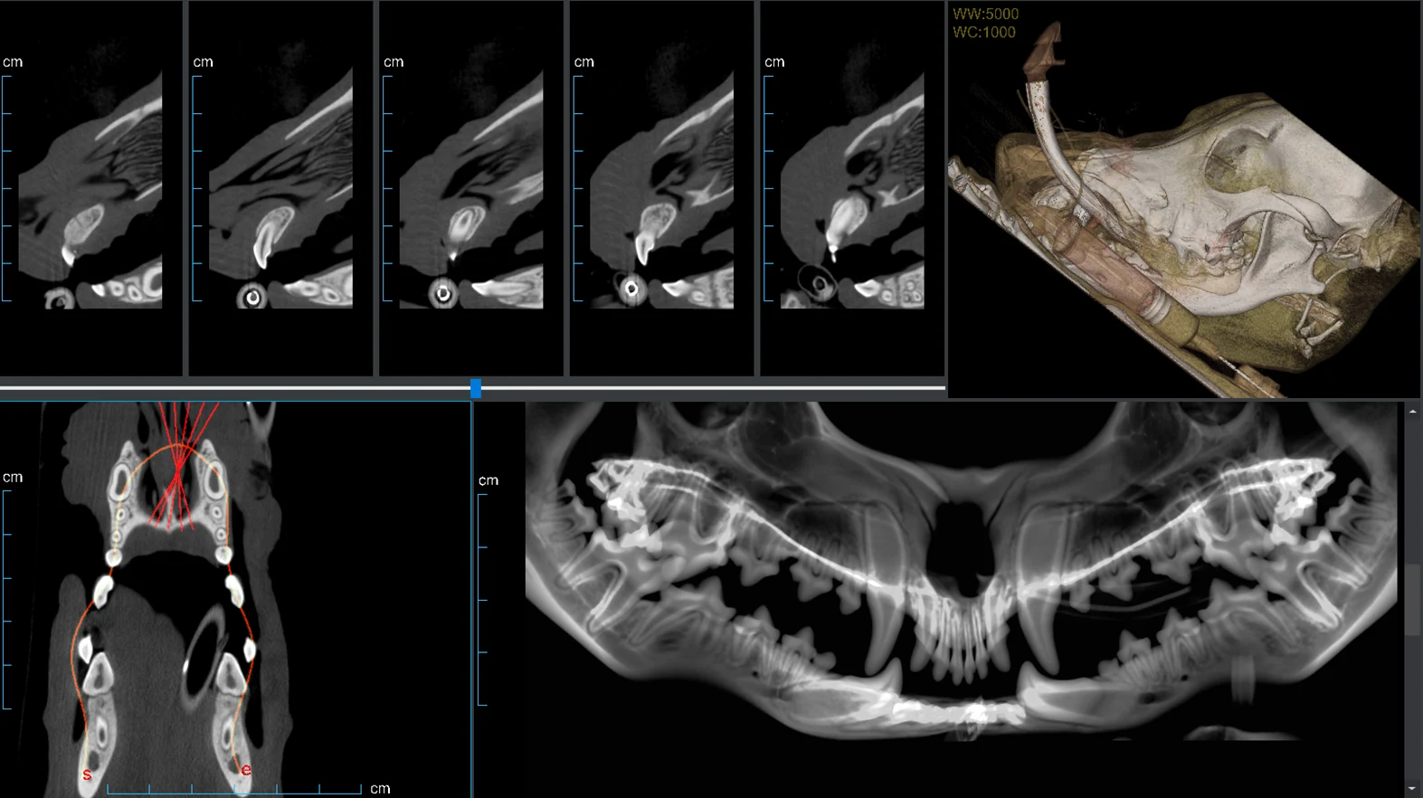
Dental imaging acquired with a veterinary CT scanner in a pet hospital.
09 · Comparison Table
Compare clinical role, image output, room requirements, workflow, training and investment before selecting a system.
| Priority | Parameter / Customer Concern | Animal CT Scanner | Veterinary DR System | Veterinary X-ray Machine |
|---|---|---|---|---|
| 1 | Imaging Dimension | 3D cross-sectional imaging | 2D digital radiography | 2D X-ray projection |
| 2 | Diagnostic Depth | Advanced diagnosis for complex cases | Routine diagnostic screening | Basic radiographic examination |
| 3 | Image Reconstruction | CT reconstruction, MPR, 3D reconstruction, volume rendering | Digital image processing | Film / CR / DR image output depending on configuration |
| 4 | Image Detail | Excellent for bone, dental, soft tissue and internal anatomy | Good for bone and routine chest / abdomen screening | Basic bone and chest / abdomen imaging |
| 5 | 3D Imaging Capability | Yes, supports 3D visualization | Limited or not available | Not available |
| 6 | Cross-Sectional Imaging | Yes | No | No |
| 7 | Best Clinical Application | Oncology, dental, orthopedic, neurology, trauma, surgical planning | Routine fracture, chest, abdomen and preoperative screening | Basic radiography and general animal examination |
| 8 | Veterinary Dental Imaging | Strong for tooth root, jaw, maxillofacial and dental abscess evaluation | Useful for dental radiography, but limited 3D information | Basic dental or skull X-ray only |
| 9 | Orthopedic Imaging | Strong for complex fractures, joints, spine, pelvis and implant planning | Good for routine fracture diagnosis | Good for basic fracture screening |
| 10 | Oncology Imaging | Suitable for tumor staging, metastasis evaluation and surgical margin planning | Limited tumor screening ability | Limited tumor screening ability |
| 11 | Neurology Imaging | Useful for skull, spine, cervical spine and selected neurological cases | Limited mainly to bone structures | Limited mainly to bone structures |
| 12 | Thoracic Imaging | Detailed lung, chest and deep lesion evaluation | Common for routine thoracic screening | Common for basic thoracic imaging |
| 13 | Abdominal Imaging | Better for internal organs and deep anatomical structures | Routine abdomen screening | Basic abdomen screening |
| 14 | Surgical Planning | Strong support for 3D surgical planning and anatomical measurement | Basic preoperative reference | Basic preoperative reference |
| 15 | Scan Speed | Fast scan acquisition, but full workflow includes anesthesia and reconstruction | Very fast digital image acquisition | Fast exposure; output speed depends on film, CR or DR setup |
| 16 | Patient Motion Requirement | Patient must remain very still | Moderate motion tolerance | Moderate motion tolerance |
| 17 | Anesthesia / Sedation Need | Often required for accurate CT imaging | Usually not required, sometimes sedation is needed | Usually not required, sometimes sedation is needed |
| 18 | Radiation Dose | Higher than single X-ray exposure; low-dose protocols are important | Lower than CT | Lower than CT |
| 19 | Low-Dose Protocol | Important feature for modern CT systems | Related to exposure control | Related to exposure control |
| 20 | Animal Compatibility | Dogs, cats, exotic animals, horses; depends on bore size, FOV and table capacity | Dogs, cats, small animals, large animals; depends on X-ray power and table design | Wide animal use; portable X-ray is common for equine applications |
| 21 | Small Animal Application | Strong for dog, cat, rabbit, bird and exotic animal CT | Excellent for routine small animal imaging | Common for basic small animal imaging |
| 22 | Equine Application | Strong for equine head, dental, sinus, limb and standing CT applications | Common for equine limb and basic radiography | Very common for portable equine X-ray |
| 23 | Bore Size / Gantry Size | Critical for animal size compatibility | Not applicable | Not applicable |
| 24 | Slice Count | Key parameter: 16-slice, 32-slice, 64-slice, 128-slice | Not applicable | Not applicable |
| 25 | Slice Thickness | Important for image resolution and small lesion detection | Not applicable | Not applicable |
| 26 | Spatial Resolution | High-resolution imaging, especially for bone and dental details | Good digital resolution depending on detector | Depends on system and imaging method |
| 27 | Detector Type | CT detector / flat panel detector depending on system type | Flat panel detector | Film, CR cassette or DR detector |
| 28 | Software Functions | MPR, 3D reconstruction, volume rendering, DICOM, PACS, measurement tools | Image processing, measurement, annotation, DICOM, PACS | Basic image output; advanced functions depend on DR/CR system |
| 29 | DICOM Compatibility | Usually supported | Usually supported in DR systems | Supported only when connected with digital imaging system |
| 30 | PACS Integration | Important for advanced hospitals and imaging centers | Common in modern DR systems | Depends on digital configuration |
| 31 | Installation Requirement | High: CT room, shielding, power supply, cooling, floor loading | Medium: X-ray room, shielding, power supply | Low to medium depending on fixed or portable design |
| 32 | Room Space Requirement | Larger space required | Smaller than CT | Smaller than CT, portable models need less space |
| 33 | Radiation Shielding | Strict shielding planning required | Shielding required | Shielding required |
| 34 | Operation Difficulty | High; requires CT workflow and image interpretation training | Medium; easier for daily clinical use | Medium; positioning and exposure experience required |
| 35 | Clinical Training Need | CT scan protocol, anesthesia workflow, reconstruction and interpretation training | Radiographic positioning and software training | X-ray positioning and exposure training |
| 36 | Maintenance Requirement | High: X-ray tube, detector, gantry, software, calibration | Medium: detector, workstation, generator, tube | Medium: tube, generator and mechanical parts |
| 37 | Equipment Cost | High investment | Medium investment | Low to medium investment |
| 38 | Return on Investment | Depends on referral cases, oncology, dental CT, orthopedic CT and high-value exams | Strong ROI through daily high-frequency imaging | Good ROI for basic clinics and mobile veterinary services |
| 39 | Daily Case Volume | Lower frequency but higher-value cases | High-frequency routine cases | High-frequency basic cases |
| 40 | Hospital Positioning | Advanced veterinary imaging center, referral hospital, specialty hospital | General animal hospital, pet clinic, veterinary practice | Basic clinic, mobile vet, equine practice |
| 41 | Main Selling Point | Advanced 3D diagnosis for complex veterinary cases | Fast daily digital radiography workflow | Affordable and flexible basic X-ray imaging |
10 · Customer Reviews
Verified customer reviews will be published here after we receive original feedback and permission from our customers.
No fabricated ratings or testimonials11 · FAQ
An animal CT scanner is a computed tomography imaging system used for veterinary diagnosis. It uses X-rays and computer reconstruction to create cross-sectional and 3D images of animal anatomy, helping veterinarians evaluate complex bone, dental, soft-tissue, thoracic, abdominal, oncology and neurology cases.
Veterinary X-ray creates a 2D projection image, while animal CT creates cross-sectional images and 3D reconstructions. CT reduces the problem of tissue overlap and provides more detailed anatomical information for complex cases.
Animal CT is better for complex diagnostic cases, surgical planning, dental CT, tumor staging, spine evaluation and detailed 3D anatomy. Veterinary DR is better for routine, fast and lower-cost daily imaging. They do not replace each other; they serve different clinical levels.
Animal CT scanners can be used for dogs, cats, rabbits, birds, exotic animals, horses and selected large animals. Actual compatibility depends on bore size, field of view, table weight capacity and patient-positioning design.
13 · Blog Listing
Five articles covering CT selection, modality comparison, anesthesia and wider imaging procurement.
Compare veterinary CT and digital X-ray by image output, clinical role, radiation planning, anesthesia, throughput, installation and total project scope.
13 min read →Veterinary CT and X-rayPlan a veterinary CT scanner purchase around patient size, clinical applications, CT type, room, radiation safety, anesthesia, workflow and service.
13 min read →Veterinary anesthesiaChoose a veterinary anesthesia machine by patient size, breathing circuit, vaporizer, ventilation, monitoring, scavenging, gas supply and service.
13 min read →Veterinary ultrasoundChoose a veterinary ultrasound machine by species, exams, probes, Doppler modes, portability, workflow, support and total cost - not headline price alone.
12 min read →Veterinary ultrasoundCompare portable, handheld and cart-based veterinary ultrasound systems by mobility, controls, probes, workflow, service and ownership cost.
11 min read →14 · Project FAQ
Selection, room planning, workflow and training should be evaluated together before a CT project is quoted.
Send an Inquiry →Yes. We do not simply recommend the most expensive system. We help customers compare animal type, case volume, clinical applications, room size, budget, expected ROI and technical skill level, then recommend a suitable imaging solution.
Yes. We can support a complete veterinary imaging solution, including an animal CT scanner, veterinary DR system, veterinary X-ray machine, ultrasound machine, anesthesia system, patient monitor, positioning accessories, image workstation and related installation guidance.
Yes. We can provide solutions for small-animal hospitals, equine clinics, referral centers, veterinary universities and imaging centers. We help customers evaluate patient size, bore size, FOV, table capacity and clinical workflow before model selection.
Yes. For CT projects, we can provide guidance on room layout, shielding requirements, power supply, air conditioning, floor loading, installation space, positioning workflow and related accessories, helping customers prepare before equipment arrival.
Yes. We can support operation training, scanning-workflow guidance, software training, 3D-reconstruction training, DICOM/PACS workflow explanation and basic application guidance for dental, orthopedic, oncology and small-animal CT cases.
15 · Warranty Period and After-Sales Service
The veterinary CT main unit and standard accessories are covered for 12 months unless the final quotation states otherwise.
Our professional sales engineers guide you and help you choose suitable animal medical equipment and project solutions.
Our professional technical engineers assist with installation and operation, online training, online upgrades, remote assistance and more.