
Short answer
Veterinary CT and digital X-ray are complementary modalities, not interchangeable price tiers. Digital radiography records projection images and is commonly the practical foundation for fast routine imaging. CT acquires many X-ray projections and reconstructs cross-sectional slices, reducing anatomical superimposition and supporting multiplanar or 3D review. CT generally requires a larger project around room design, radiation protection, patient immobilization or anesthesia, reconstruction software, staffing and service. The correct modality is the least complex system that answers the clinic's defined clinical questions reliably and safely.
Editorial note: This independent procurement guide was prepared from Mevidon's supplied product materials and the primary professional or government sources listed below. It contains no paid rankings and does not replace clinical, engineering or regulatory advice.
Quick comparison
| Project factor | Veterinary X-ray / DR | Veterinary CT |
|---|---|---|
| Image output | 2D projection radiographs | Cross-sectional slices and reconstructions |
| Common role | Fast routine and first-line imaging | Detailed targeted or referral imaging |
| Patient workflow | Positioning; often brief acquisition | Protocol, immobilization and sometimes anesthesia |
| Facility scope | Radiography room or mobile workflow | Dedicated scanner, control and support planning |
| Data volume | Relatively smaller studies | Larger series requiring review and storage |
How do CT and X-ray create different images?
Digital radiography sends an X-ray beam through the patient and records a two-dimensional projection on a detector. Structures along the beam path overlap. CT records many projections around the patient and uses computer reconstruction to create cross-sectional slices. The dataset can be viewed in different planes and may support 3D reconstructions. Both use ionizing radiation; ultrasound and MRI do not.
The difference in image structure changes workflow and interpretation. CT is not simply a sharper radiograph, and a radiograph is not a low-cost CT slice. The veterinarian should select the modality that addresses the clinical question and patient condition.
When is digital X-ray the practical starting point?
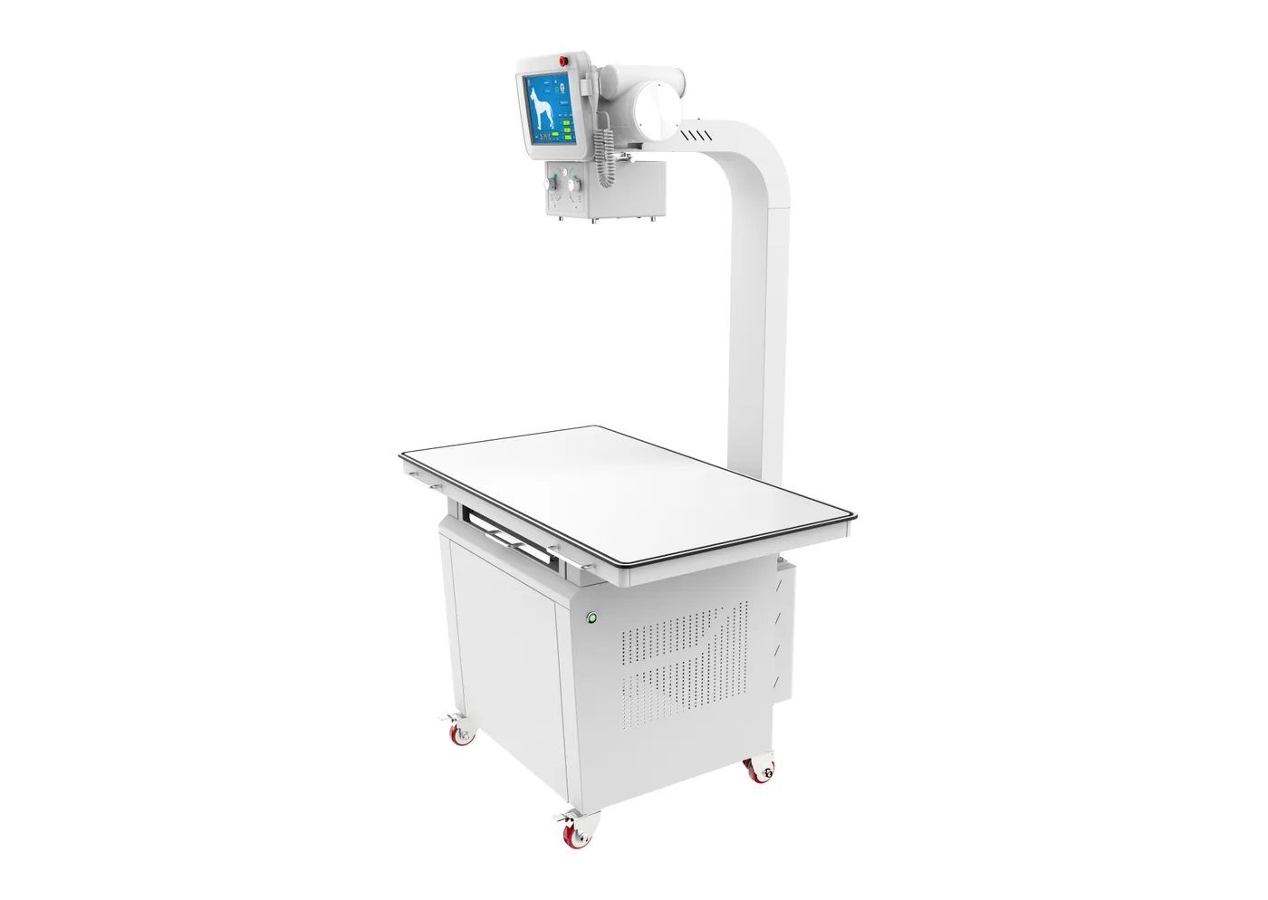
Digital radiography is often the first project for general practices needing rapid thoracic, abdominal or skeletal projection imaging. It supports a broad case mix, relatively fast acquisition and familiar interpretation workflows. Portable or mobile configurations may extend use, subject to local radiation rules. The procurement brief should define generator power, detector size, table or stand, positioning accessories, acquisition software and storage.
Radiographs may be limited by anatomical superimposition and positioning. Multiple views can help, but some questions still require cross-sectional imaging, ultrasound, MRI or referral. Equipment selection should follow the service scope rather than an assumption that one modality replaces all others.
When does a veterinary CT project make sense?
CT becomes relevant when the service needs cross-sectional detail, reduced superimposition, multiplanar review, 3D planning or a referral-level imaging workflow. Potential applications vary by scanner design, patient size, anatomy and protocol. A business case should identify expected referrals, case volume, reporting expertise and how CT findings will change treatment or surgical planning.
CT acquisition may require deep sedation, anesthesia or another immobilization plan, depending on the patient, region and scanner speed. The clinic must design this pathway with a qualified veterinarian or anesthesiologist and include airway access, monitoring, transfer and recovery - not treat patient management as an accessory.
What are the radiation-protection implications?
Both modalities use ionizing radiation. FDA guidance emphasizes justification - performing an exam when it is needed to answer a medical question - and optimization, using the lowest exposure that yields adequate diagnostic information. IAEA Safety Reports Series No. 104 applies radiation-protection principles specifically to veterinary medicine and addresses workers and the public.
Before purchase, involve the local regulator, a qualified radiation-protection professional and the facility team. Shielding, controlled areas, warning systems, operating procedures, staff training, quality assurance and registration requirements vary by jurisdiction. A supplier's room drawing does not replace a locally approved design.
How do room and infrastructure requirements compare?
A DR project may include the generator, tube stand, table or wall stand, detector, workstation and a protected room or compliant mobile workflow. A CT project adds gantry and patient-table geometry, control area, cooling or environmental limits, higher data transfer and storage, service access, patient preparation and anesthesia or contrast zones. Power quality and backup arrangements also need confirmation.
Ask for a formal pre-installation document with equipment dimensions, weights, heat load, electrical characteristics, network requirements, access route and service clearances. Verify these details against the final model before construction begins.
How do throughput, staffing and interpretation differ?
Radiography usually supports rapid positioning, exposure and review, while CT adds patient preparation, protocol selection, positioning, scan and reconstruction, larger datasets and often recovery. CT throughput therefore depends on more than scan time. Staff must be trained in patient handling, radiation safety, acquisition protocols, contrast procedures where applicable, image reconstruction and emergency response.
Interpretation capacity belongs in the business case. Define whether studies are read internally or through teleradiology, required turnaround times, DICOM routing, internet reliability and how urgent cases are escalated. A scanner without a dependable reporting workflow cannot deliver its intended value.
How should total project cost be compared?
Compare installed and operational scope, not main-unit price. Include shielding design, construction, power, HVAC, network, workstation, injector or positioning accessories, anesthesia and monitoring integration, shipping, customs, installation, acceptance testing, training, software, storage, maintenance, tubes or detectors and service travel. Model expected case volume and downtime.
Use the same requirement document for each supplier. Separate included equipment, optional items, local contractor work and responsibilities after delivery. For many clinics, DR plus reliable referral access is a stronger first step than underused CT; for a mature referral service, CT may unlock cases that projection imaging cannot adequately resolve.
Copy this buyer checklist
- Which clinical questions and anatomy must the system address?
- What patient sizes must fit safely?
- Is the project routine radiography, referral imaging or both?
- Who will approve shielding, controlled areas and local registration?
- How will immobilization, anesthesia, monitoring, contrast and recovery be handled?
- Who interprets studies and how are DICOM files transferred?
- What installation, uptime, training and service scope is included?
Buyer FAQs
Is CT better than X-ray for every veterinary case?
No. CT offers cross-sectional detail, while radiography is fast and valuable for many routine questions. The veterinarian should choose the modality that appropriately answers the clinical question.
Do veterinary CT scanners use radiation?
Yes. CT and conventional radiography use ionizing X-rays. Facilities need justified protocols, optimized exposure, trained staff and radiation-protection arrangements that meet local requirements.
Does every animal need anesthesia for CT?
No universal rule applies. Immobilization needs depend on the patient, anatomy, protocol, scan speed and clinical judgment. The responsible veterinary team must design the sedation or anesthesia pathway.
Can a clinic replace X-ray with CT?
Usually the modalities are complementary. A clinic should assess routine workload, referral access, room constraints, interpretation capacity and ownership cost before changing its imaging mix.
Sources and further reading
Primary sources were accessed and checked on July 19, 2026.
- FDA: Medical X-ray ImagingDefines radiography and CT, and explains justification, optimization and ALARA principles.
- FDA: Computed Tomography (CT)Explains cross-sectional CT imaging, benefits, radiation considerations and facility responsibilities.
- IAEA Safety Reports Series No. 104Veterinary-specific guidance for worker and public radiation protection, facility planning and safe operation.
Important procurement note
This guide is educational and does not replace clinical, engineering, radiation-safety, occupational-safety, regulatory or facility advice. Product capability and compliance depend on the final model and market. Confirm the signed specification, included accessories, local requirements, installation scope and service plan before purchase.