
Short answer
The best veterinary ultrasound machine is the system that reliably covers your most common species and examinations with the right probes, imaging modes and workflow. Begin with a written clinical use profile, then compare probe compatibility, image controls, portability, data export, training and service. A lower purchase price can become expensive if a required probe, battery, trolley, DICOM option or local support is excluded. Ultrasound uses non-ionizing energy, but safe and useful examinations still depend on trained operators, appropriate settings and a medically justified workflow.
Editorial note: This independent procurement guide was prepared from Mevidon's supplied product materials and the primary professional or government sources listed below. It contains no paid rankings and does not replace clinical, engineering or regulatory advice.
Quick comparison
| Decision | Portable laptop | Handheld / wireless | Cart-based |
|---|---|---|---|
| Best fit | Mixed routine imaging | Rapid point-of-care checks | High-volume or advanced imaging |
| Mobility | High | Very high | Low to moderate |
| Controls | Dedicated keyboard and trackball | Touch or mobile interface | Large control surface |
| Probe access | Broad; model dependent | Usually probe-specific | Often broadest; model dependent |
| Primary tradeoff | Balance of performance and transport | Fast access versus smaller interface | Workflow and capability versus space |
What should you decide before comparing machines?
Write a one-page use profile before requesting quotations. Record the species you treat, the five examinations that generate most of the workload, expected weekly volume, scanning locations and current operator skill. This converts a broad search for a veterinary ultrasound machine into a measurable specification. A companion-animal clinic doing routine abdominal scans needs a different package from an equine reproductive service, a cardiology referral center or a mobile mixed practice.
Also separate today's essential work from likely expansion during the next two or three years. Buying every optional feature immediately can waste budget, while choosing a closed system with no practical upgrade route can force premature replacement. Ask suppliers to label each capability as included, optional, license-based or unavailable.
- Species and typical patient weight range
- Abdominal, cardiac, reproductive, vascular, superficial or musculoskeletal exams
- Clinic room, operating room, farm call or mobile service
- Current users, training needs and expected case volume
Which ultrasound probes do you need?
Probe selection connects the scanner to the examination. A micro-convex or convex probe is commonly considered for general abdominal work; a linear probe supports superficial structures and vascular access; a phased-array probe provides a smaller acoustic window for cardiac work; and transrectal formats may be required for large-animal reproductive examinations. These are starting points, not universal prescriptions. Frequency range, footprint, penetration, cable length and ergonomics must match species and anatomy.
Ask for the exact probe model numbers and quantities in the signed quotation. Confirm which ports can remain active, whether adapters are required, how probes are cleaned, replacement lead time and warranty exclusions. A capable console with the wrong probe package is not a usable solution.
Which imaging modes are worth paying for?
B-mode is the foundation for anatomical imaging. M-mode is used for motion-based assessment, while color and power Doppler visualize flow and spectral Doppler supports velocity analysis. Tissue Doppler, elastography, contrast imaging, 3D or 4D should be justified by a repeatable clinical or teaching workflow. Do not treat a long mode list as proof of image quality or ease of use.
During a demonstration, ask the supplier to show the relevant mode on a comparable species and anatomy, then let your intended operator adjust depth, gain, focus, dynamic range, Doppler scale and presets. Save and export representative studies. The value lies in reproducible images and efficient controls, not in a feature name on a brochure.
Portable, handheld or cart-based: which form factor fits?
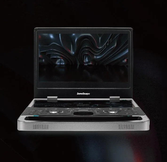
Choose form factor from the work environment. Laptop-style systems balance mobility with dedicated controls and multiple applications. Handheld or wireless systems can shorten the path to rapid point-of-care checks, field work and crowded rooms, but screen size, battery dependence and probe-specific design may limit longer studies. Cart-based systems provide a larger display, ergonomic controls and often more probe or connectivity options, but require floor space and room-to-room access.
Image capability is model-specific; portability alone does not determine image quality. Compare systems using the same exam task, probe class and operator whenever possible. Include trolley stability, carrying case, charging method, boot time and disinfection workflow in the trial.
How should image quality be evaluated?
A live demonstration is more useful than a single polished sample image. Prepare a repeatable test plan covering the anatomy and patient size that matter to your service. Evaluate penetration, contrast, spatial detail, uniformity, motion response, Doppler sensitivity and artifact behavior, then record how many adjustments the operator needs to reach a diagnostic view. Ask whether presets can be saved by species, exam and user.
FDA information describes ultrasound as non-ionizing but still recommends prudent, medically appropriate use by trained personnel. Procurement should therefore include application training, operator documentation and access to service - not only the console and probes. Mevidon can coordinate configuration comparisons, while the responsible veterinarian determines clinical protocols.
Which workflow and connectivity details are easy to miss?
Check boot time, battery duration under a stated test condition, active probe ports, patient database, reporting templates, language, image and video export, USB or network transfer, DICOM capability, printer support and user access controls. Confirm storage capacity and how studies are backed up. If the clinic uses a PACS or practice-management system, request a workflow test rather than accepting a generic compatibility claim.
Cleaning and transport also affect uptime. Review probe-care instructions, fan and filter access, splash resistance statements, trolley dimensions and the availability of replacement batteries, power adapters and common accessories. Document who owns integration work and any recurring software fees.
How do you compare total cost and supplier support?
Build a three-to-five-year cost sheet. Include the main unit, every probe, trolley or carrying case, printer, DICOM or measurement packages, shipping, taxes, installation, training, warranty extensions, planned maintenance and likely replacement accessories. Record what happens if a probe fails and whether remote diagnosis, loan equipment or local engineers are available in your market.
Use the same written scope for every supplier. Mevidon quotations should identify source-supported specifications, included items, optional items and points that still require confirmation. Final compliance documents, delivery time and service terms depend on the model and destination market and must be confirmed before purchase.
Copy this buyer checklist
- Which species, anatomy and examinations make up at least 80% of the workload?
- Which probes and Doppler modes must be included on day one?
- Does the system need DICOM, reporting, printer or network connectivity?
- What battery, trolley, transport and cleaning accessories are required?
- Can the intended operator complete a comparable live demonstration?
- Who provides training, warranty coordination and replacement probes?
- What is the complete three-to-five-year ownership cost?
Buyer FAQs
How much does a veterinary ultrasound machine cost?
There is no useful single price because probe count, Doppler capability, form factor, software, accessories, shipping and service scope change the quotation. Compare complete configurations for the same exam profile instead of bare-console prices.
Is a portable veterinary ultrasound good enough for a clinic?
Often yes for routine and mobile workflows, provided the model, probes and imaging modes match the exams. A live task-based demonstration is the best way to verify performance and ergonomics.
Which probe is best for veterinary ultrasound?
No one probe covers every exam. Convex or micro-convex, linear, phased-array and transrectal probes serve different anatomy, depth and access needs. The package should follow the clinic's species and exam mix.
Does ultrasound use ionizing radiation?
No. Diagnostic ultrasound uses high-frequency sound waves rather than ionizing radiation. It should still be used prudently by trained professionals for a medically appropriate purpose.
Sources and further reading
Primary sources were accessed and checked on July 19, 2026.
- FDA: Ultrasound ImagingExplains real-time ultrasound imaging, non-ionizing energy, benefits, risks and prudent use.
- Mevidon veterinary ultrasound catalogSource-supported portable, handheld, cart-based and species-oriented configurations used in this procurement framework.
Important procurement note
This guide is educational and does not replace clinical, engineering, radiation-safety, occupational-safety, regulatory or facility advice. Product capability and compliance depend on the final model and market. Confirm the signed specification, included accessories, local requirements, installation scope and service plan before purchase.