
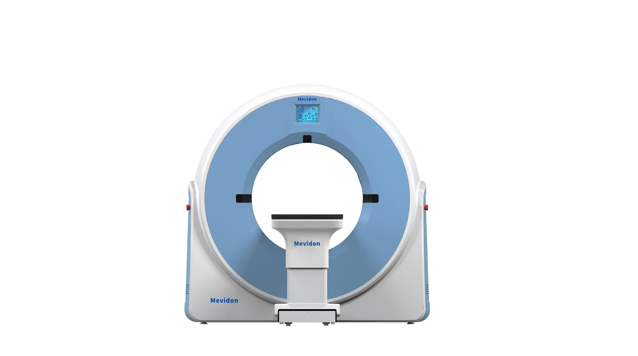
The right veterinary CT scanner is the system that fits your patients, clinical services, room conditions, staff capabilities, expected workload, and long-term budget. Begin with the examinations you need to perform, then verify geometry, image evidence, anesthesia integration, facility planning, connectivity, service, and total ownership cost.
Investing in a veterinary CT scanner is not simply an equipment purchase. It is a clinical, operational, financial, and facility-level decision that can change how an animal hospital diagnoses complex disease, plans surgery, receives referral cases, and develops specialist services.
Computed tomography uses X-rays and computer reconstruction to create cross-sectional images. Compared with conventional radiography, CT reduces the problem of overlapping anatomy and can provide more detailed information about the skull, spine, lungs, joints, teeth, nasal cavity, and internal organs. The American College of Veterinary Radiology explains the CT process and the need for conscientious radiation-safety practices. The best system is not automatically the model with the highest slice count, the largest price tag, or the longest feature list.
Review image evidence from the cases you expect to scan.
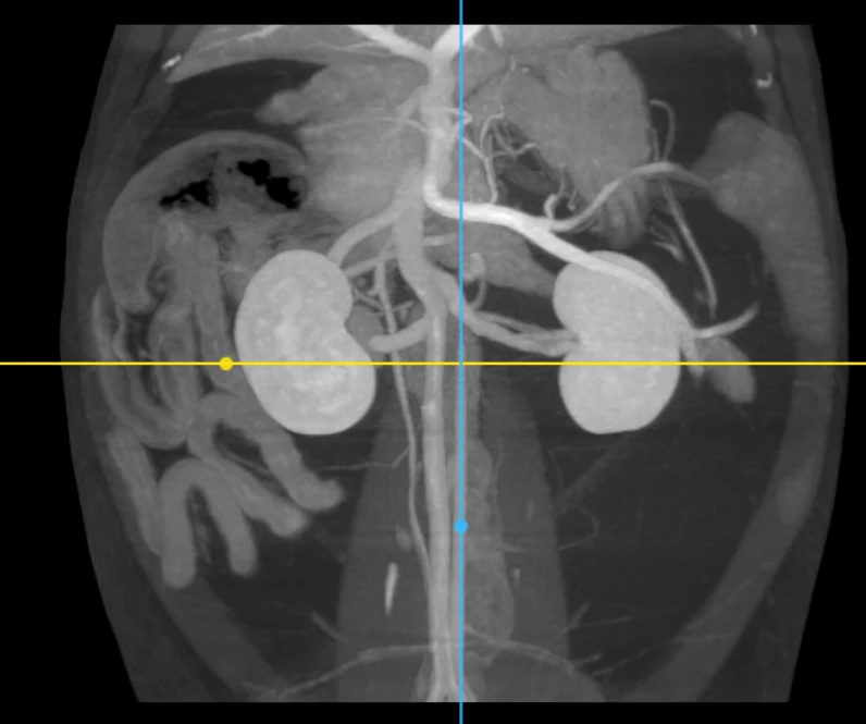
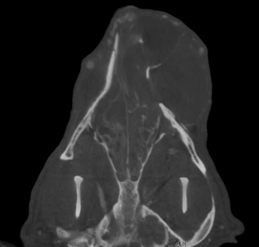
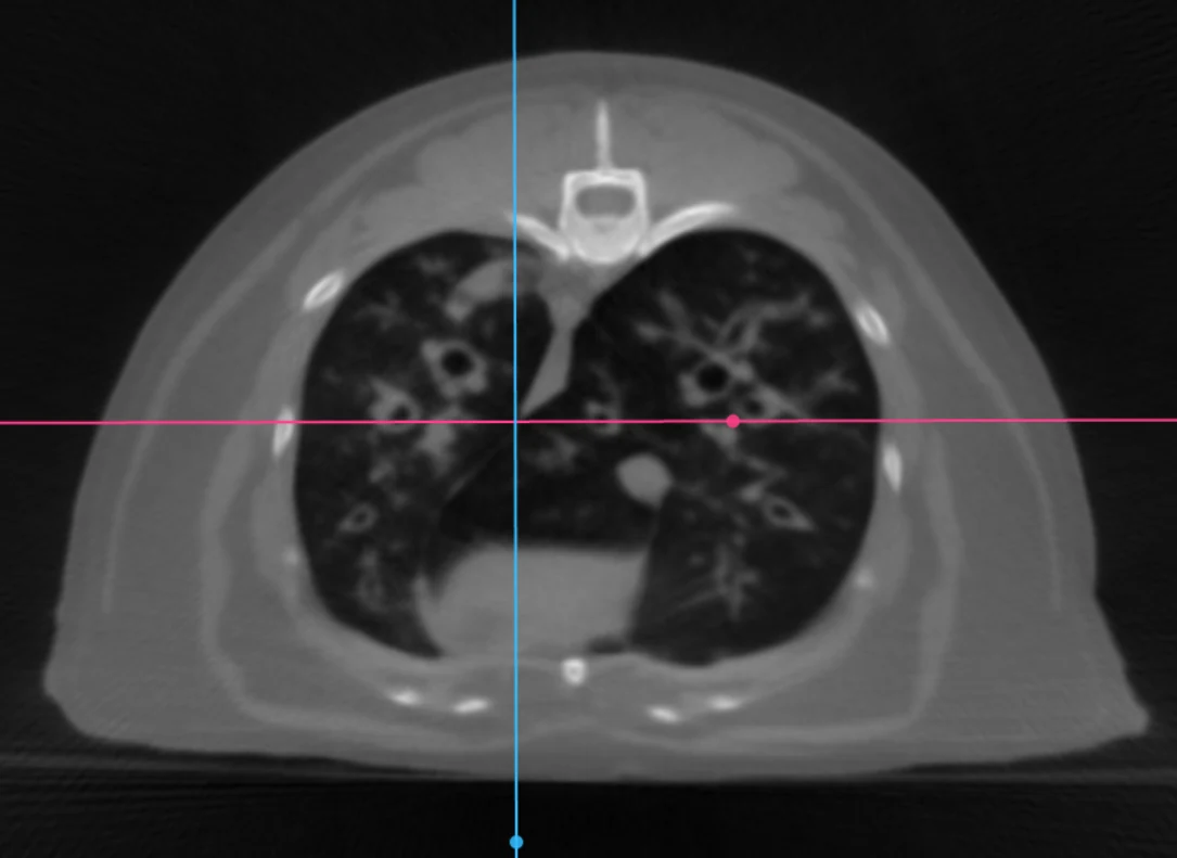
Supplied case images are presented as imaging references for equipment and workflow evaluation; they are not patient-specific diagnostic advice.
Veterinary CT imaging cases
Use the supplied case reference alongside complete anonymized DICOM studies when evaluating clinical image quality.
Start with the Clinical Problems You Need to Solve
Before discussing technical specifications, review the cases your hospital currently handles and the cases you want to attract. A general small-animal hospital may use CT for skull and nasal disease, dental and ear evaluation, complex fractures, spinal cases, thoracic imaging, tumor staging, trauma, and surgical planning. A referral hospital may also require advanced oncology, neurology, orthopedics, CT angiography, radiation planning, or image-guided workflows. An equine practice may focus on the head, teeth, sinuses, or distal limbs.
Create a list of your ten most important examinations, expected monthly case volume, smallest and largest patients, and the clinical decisions each scan must support. This is more useful than beginning with, “Do we need 32 slices or 64 slices?”
Match Scanner Geometry to Your Animal Population
Veterinary patients vary widely in size and body shape. A scanner suitable for cats, rabbits, and small dogs may not accommodate giant-breed dogs, large exotic animals, or horses. Ask the supplier to confirm the gantry aperture, usable field of view, table width, table load, scannable body length, and clearance for anesthesia tubing and monitoring cables.
Do not rely only on the published bore diameter. A large opening does not guarantee that the shoulders, thorax, head, or limbs can be positioned correctly. Send the supplier representative patient measurements and intended positions. Request written confirmation, layout drawings, and details of available positioning aids. For equine projects, confirm whether the system is designed for recumbent scanning under anesthesia, standing examinations, or only specific anatomical regions.
Compare Conventional CT and Cone-Beam CT
Veterinary buyers often compare conventional multidetector CT with cone-beam CT, or CBCT. Conventional CT is generally chosen for broad clinical flexibility, fast acquisition, whole-body imaging, soft-tissue assessment, thoracic studies, oncology, trauma, and contrast examinations. CBCT uses a cone-shaped X-ray beam and flat-panel detector. It can be attractive for dental, maxillofacial, orthopedic, and other high-detail bone applications, and some systems offer a compact footprint.
Soft-tissue contrast, motion sensitivity, field of view, artifacts, and scan time vary considerably between models. Do not choose by the words “CT” or “CBCT” alone. Request anonymized DICOM studies from similar animals and the same anatomical regions you plan to examine. Ask a veterinarian or radiologist to judge whether the images answer your clinical questions.
Understand Slice Count Before Comparing Prices
Slice count is heavily marketed, but it should never be evaluated alone. Multi-slice CT uses multiple detector rows to acquire several slices during one gantry rotation. Wider coverage and faster acquisition may improve workflow and reduce motion problems, while thin-slice acquisition supports multiplanar and 3D reconstruction. Detector configuration, z-axis coverage, rotation time, reconstruction method, and spatial resolution all contribute to performance.
Ask every supplier to state these values separately
- Physical detector rows
- Acquired slices per rotation
- Reconstructed or displayed slices
- Minimum acquired and reconstructed slice thickness
- Rotation time and scan speed
- Spatial and contrast resolution
- Reconstruction time
A higher reconstructed slice number does not necessarily mean that the scanner physically acquires the same number of independent slices. Ask the supplier to explain the difference in writing. For many hospitals, reliable protocols, suitable coverage, consistent image quality, and affordable service are more valuable than the highest available slice count.
Judge Image Quality with Real Veterinary Cases
A brochure may contain excellent images acquired with an ideal protocol or a different configuration. Request complete sample studies for the anatomy most relevant to your hospital: skull, nasal cavity, ear, teeth, spine, joints, thorax, abdomen, tumors, and any equine region you expect to scan. Review edge definition, image noise, soft-tissue differentiation, metal artifacts, motion artifacts, and the quality of multiplanar reconstruction.
Check whether thin slices remain clinically useful rather than becoming excessively noisy. Ask whether the proposed configuration includes iterative reconstruction, metal-artifact reduction, automatic exposure control, adjustable low-dose protocols, and application packages for dental, orthopedic, vascular, or oncology imaging. Do not pay for advanced software before confirming that your team will use it.
Include Anesthesia and Monitoring in the Project
Many veterinary CT studies require deep sedation or general anesthesia to prevent movement. Anesthesia planning is therefore part of the CT project, not an optional accessory. A complete workflow may require a veterinary anesthesia machine, oxygen supply, suitable breathing circuits, multiparameter monitoring, capnography, pulse oximetry, blood-pressure measurement, ECG, infusion equipment, long monitoring lines, emergency supplies, and a safe recovery area.
The monitoring display should remain visible to staff, while cables and tubing must not interfere with table movement. Your team should have written procedures for induction, positioning, contrast administration, ventilation when needed, monitoring, recovery, and emergency access. Fast acquisition can shorten time in the gantry, but total anesthesia time also includes preparation, positioning, contrast studies, repeat scans, and recovery. Final protocols remain the responsibility of qualified veterinary professionals.
Verify Radiation Safety and Room Requirements Early
A veterinary CT scanner usually requires more than an empty room and a suitable power outlet. Facility planning may include a dedicated CT room, shielding calculations, a control area, access control, warning systems, electrical capacity, grounding, cooling, floor loading, delivery access, service clearances, network connections, and space for anesthesia and monitoring equipment.
The International Atomic Energy Agency’s veterinary radiation-safety guidance covers facility design and protection of workers and the public. Before ordering, request the pre-installation document, equipment dimensions, heat output, electrical requirements, service clearances, and shielding information for the exact model. Local regulations differ, so general supplier advice must be reviewed by qualified local experts and authorities. Compact or self-shielded designs may reduce construction work in some projects, but they do not remove the need for a formal local radiation assessment.
Examine Software, DICOM, PACS, and Reporting
CT creates many images, so the scanner must fit your hospital’s information system. Confirm support for DICOM storage and transfer, PACS integration, original DICOM export, patient worklist where required, multiplanar reconstruction, volume rendering, measurement tools, protocol customization, data backup, and teleradiology.
DICOM is the international standard used to produce, store, display, send, query, process, and retrieve medical imaging information. Ask the supplier to demonstrate the full process: patient registration, protocol selection, acquisition, reconstruction, export, PACS transfer, and remote reporting. Excellent images provide limited value if staff cannot move the study efficiently or share full data with a radiologist.
Calculate Total Cost of Ownership
The scanner price is only one part of the investment. Your budget should also include shipping, customs, room renovation, shielding, electrical and cooling upgrades, installation, anesthesia and monitoring equipment, workstations, PACS storage, staff training, preventive maintenance, software licenses, replacement parts, and service travel.
Ask for a five-year ownership estimate. Clarify X-ray tube warranty, detector coverage, maintenance intervals, remote diagnostics, spare-parts availability, response time, and the process for major repairs. A lower purchase price can become expensive when parts are unavailable, service is slow, or the hospital repeatedly cancels examinations because of downtime.
Evaluate Training and Supplier Capability
CT performance depends on hardware, software, protocols, and people. Your team must learn patient positioning, anesthesia coordination, contrast timing, scan selection, reconstruction, artifact recognition, quality control, data transfer, and basic troubleshooting.
Request a written training and support plan covering operator training, veterinary applications, protocol setup, routine calibration, software use, DICOM/PACS, remote assistance, and refresher training. Also request an installation responsibility matrix showing who handles room preparation, unloading, positioning, electrical connection, acceptance testing, application training, and regulatory documentation. A capable supplier should ask detailed questions about your patients, clinical goals, room, staff, and service expectations before recommending a model.
Choose a complete CT solution
Make the final decision around the entire clinical project.
The right veterinary CT scanner should improve diagnostic confidence, support treatment planning, fit your animal population, and operate reliably within your hospital’s workflow. Avoid buying from a brochure alone. Request real case images, technical documents, room requirements, a complete configuration list, and a written service plan. Ideally, involve clinical users, anesthesia staff, management, IT personnel, facility engineers, and radiation-safety professionals in the final decision.
Animal hospitals, referral centers, veterinary universities, and imaging projects can explore the Mevidon Veterinary CT Scanner collection, including small-animal, equine, dental, cone-beam, orthopedic, and oncology-oriented CT directions. Mevidon can also help buyers compare patient size, applications, room conditions, anesthesia workflow, related imaging equipment, and the final project configuration before quotation.
Technical references
Procurement and facility decisions should be checked against the selected model’s final technical documentation and applicable local regulations.